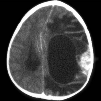
Computed tomography
In diagnostic imaging, computed tomography (also known as CT scan or CAT scan) is a form of tomography that uses a computer algorithm to reconstruct the image.
Methods of creating the data for tomographic imaging differ on whether the image is constructed by sending energy generated from an external source through the target, or if the energy comes from a source internal to the target (typically a radionuclide injected into the target's metabolic processes).
In both cases, the receiver(s) for the signal, as modified by the target, are typically mounted on a gantry that rotate them around the target, and then moves the gantry in small distances along the length of the target.
Classification
- X-ray computed tomography. This is defined as "tomography using x-ray transmission and a computer algorithm to reconstruct the image."[1] It is generally considered a part of diagnostic radiology, although many specialists, especially trauma surgeons, are adept at quick interpretation of scans.
- Spiral computed tomography has a set of concurrently operating detectors at different angles of view. Examples include computed tomographic pulmonary angiography for detecting pulmonary embolism, computed tomographic cardiac angiography for detecting coronary heart disease[2], and computed tomographic colonography (virtual colonoscopy).
- Cone CT is uses a cone or pyramid-shaped beam of radiation.[3].
- Emission Computed Tomography This is defined as "tomography using radioactive emissions from injected radionuclides and computer algorithms to reconstruct an image".[4] Formally, it is considered a part of diagnostic nuclear medicine, although it may be performed by other specialties.
- Positron emission tomography (PET Scan) is defined as "an imaging technique using compounds labeled with short-lived positron-emitting radionuclides (such as carbon-11, nitrogen-13, oxygen-15 and fluorine-18) to measure cell metabolism. It has been useful in study of soft tissues such as cancer; cardiovascular system; and brain. SPECT is closely related to PET, but uses isotopes with longer half-lives and resolution is lower."[5] An example is 18-Fluorodeoxyglucose (FDG) positron emission tomography for localizing some cancers.
- Single-Photon Emission-Computed Tomography (SPECT) is defined as "A method of computed tomography that uses radionuclides which emit a single photon of a given energy. The camera is rotated 180 or 360 degrees around the patient to capture images at multiple positions along the arc. The computer is then used to reconstruct the transaxial, sagittal, and coronal images from the 3-dimensional distribution of radionuclides in the organ. The advantages of SPECT are that it can be used to observe biochemical and physiological processes as well as size and volume of the organ. The disadvantage is that, unlike positron-emission tomography where the positron-electron annihilation results in the emission of 2 photons at 180 degrees from each other, SPECT requires physical collimation to line up the photons, which results in the loss of many available photons and hence degrades the image".[6]
Adverse effects
The risk associated with a CT scan (the increased risk of cancer associated with the radiation doses) is extremely low for any one person. However, given the increasing number of CT scans being obtained, the increasing exposure to radiation in the population may be a public health issue in the future. [7]
References
- ↑ National Library of Medicine. Tomography, X-Ray Computed. Retrieved on 2007-12-09.
- ↑ Stein PD, Yaekoub AY, Matta F, Sostman HD (August 2008). "64-slice CT for diagnosis of coronary artery disease: a systematic review". The American journal of medicine 121 (8): 715–25. DOI:10.1016/j.amjmed.2008.02.039. PMID 18691486. Research Blogging.
- ↑ Anonymous (2025), Cone-Beam Computed Tomography (English). Medical Subject Headings. U.S. National Library of Medicine.
- ↑ National Library of Medicine. Tomography, Emission-Computed. Retrieved on 2007-12-09.
- ↑ Positron-Emission Tomography. Retrieved on 2007-12-09.
- ↑ National Library of Medicine. Tomography, Emission-Computed, Single-Photon. Retrieved on 2007-12-09.
- ↑ Brenner DJ, Hall EJ (2007). "Computed tomography--an increasing source of radiation exposure". N Engl J Med 357: 2277–84. DOI:10.1056/NEJMra072149. PMID 18046031. Research Blogging.
