Flash radiography: Difference between revisions
imported>Howard C. Berkowitz No edit summary |
imported>Howard C. Berkowitz No edit summary |
||
| Line 1: | Line 1: | ||
{{subpages}} | |||
{{TOC|right}} | |||
'''Flash radiography''' is a technique to photograph objects moving at extremely high speed, such as in explosions, to allow the measurement of "their position, speed, shape, and internal density profiles...radiographic images are used to validate computer models and to experimentally determine material parameters in transient interactions, including high temperature and pressure regimes." While it has [[dual-use]] civilian applications, it is a technology critical to the development of [[nuclear weapon]]s and advanced conventional [[explosives]]. | '''Flash radiography''' is a technique to photograph objects moving at extremely high speed, such as in explosions, to allow the measurement of "their position, speed, shape, and internal density profiles...radiographic images are used to validate computer models and to experimentally determine material parameters in transient interactions, including high temperature and pressure regimes." While it has [[dual-use]] civilian applications, it is a technology critical to the development of [[nuclear weapon]]s and advanced conventional [[explosives]]. | ||
Any flash radiography system used to image explosions has the main components: | |||
#Illumination source (X-rays or protons) | |||
#an experimental configuration which contains the blast and debris from dynamic experiments | |||
#a high resolution and quantum efficiency detection system to record the radiographic image | |||
#analysis software to interpret the image into physically useful information.<ref>{{citation | |||
| url = http://www.dtic.mil/mctl/MCTL/Sec15MCTLg.pdf | | url = http://www.dtic.mil/mctl/MCTL/Sec15MCTLg.pdf | ||
| title = Militarily Critical Technologies List | | title = Militarily Critical Technologies List | ||
Revision as of 20:29, 17 May 2010
Flash radiography is a technique to photograph objects moving at extremely high speed, such as in explosions, to allow the measurement of "their position, speed, shape, and internal density profiles...radiographic images are used to validate computer models and to experimentally determine material parameters in transient interactions, including high temperature and pressure regimes." While it has dual-use civilian applications, it is a technology critical to the development of nuclear weapons and advanced conventional explosives.
Any flash radiography system used to image explosions has the main components:
- Illumination source (X-rays or protons)
- an experimental configuration which contains the blast and debris from dynamic experiments
- a high resolution and quantum efficiency detection system to record the radiographic image
- analysis software to interpret the image into physically useful information.[1]
X-ray
There are several ways to produce the X-rays:
| Generator technology | Energy | Notes |
|---|---|---|
| Linear induction accelerators (LIAs | 20 MeV single pulse, 500 R dose at 1 m in 60 ns from ~ 2-mm spot. | |
| Pulsed power accelerators | 14 MeV ~ 100 kA | Larger spot than LIA |
| High intensity laser beams | 100 MeV | order of ten micron spot size |
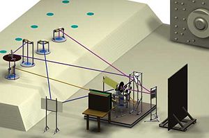
A computer-aided design layout shows placement of the experiment, which is surrounded by mirrors on stands, flashlamps, and a radiographic film cassette. Extremely energetic x rays emerge from the Flash X Ray machine through a port (upper right). Behind the experiment is the protective enclosure for radiographic film. Images from a series of mirrors are reflected to four optical cameras, providing different viewpoints. Each optical path is indicated by a different color. The cameras are located directly below the four ports on the left.[2]
In the late 1990s, the flash source at Lawrence Livermore National Laboratory was a induction linear accelerator. A gamma ray camera, rather than traditional X-ray film, captures the image.
An injector introduces an electron beam into the FXR accelerator. After passing through the accelerator, the beam enters a drift section that directs it toward a 1-millimeter-thick strip of tantalum, called a target. As the high-energy electrons pass through the target, the electric field created by the stationary charged particles of the heavy tantalum nuclei causes the electrons to decelerate and radiate some of their energy in the form of x rays. The product of this slowing process is called bremsstrahlung (braking) radiation. The x-ray photons travel toward the exploding device, where most are absorbed. The photons that make it to the camera are the image data.[3]
Proton beam
Proton radiography (pRad) utilizes conventional high energy accelerator technology to provide bursts of higher kinetic energy (1–50 GeV) protons distributed uniformly across objects to create flash radiographs of equivalent mass-profile information to the best flash X-ray radiographs
References
- ↑ Under Secretary of Defense, Acquisition, Technology and Logistics Pentagon, VA (June 2009), Section 15, Nuclear Systems Technology; Section 15.1, Flash Radiography, Militarily Critical Technologies List, U.S. Department of Defense
- ↑ "Ramrods Shepherd Hydrodynamic Tests", Science and Technology Review, Lawrence Livermore National Laboratory, September 2007
- ↑ Katie Walker (May 1997), Better Flash Photography using the FXR, Lawrence Livermore National Laboratory
